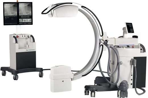
Description
OrthoScan FD mini c-arm with a flat detector. This flat detector design and compact form factor allows optimal positioning in the surgical environment. Exceptional image quality by increased grayscale improves diagnostic accuracy. User-driven design allows optimal image quality with minimal effort. Surgical lights on the undersurface of the x-ray source reduce shadows by providing primary light in the surgical field.
Thinnest Flat Detector
The FD-OR utilizes the thinnest flat detector housing on the market, allowing the user to place the detector directly on the operating surface and leave the c-arm in the field to reduce OR time and decrease sterility risks. The compact form factor allows for additional maneuverability during cases and improves workflow.
Over-Rotation
The OrthoScan mini c-arm design allows for an orbital rotation of 150°. This enables the user to take advantage of the larger field of view without making mechanical adjustments required by competitive equipment. The increased orbital movement enables preferred views without stressing anatomy.
Bilateral Sterile Field Controls
Conveniently located on each side of the tube head assembly, bilateral controls provide easy access to imaging and documentation in the sterile field.
Greater arc depth of 19.2
The “live” image is 38% larger than our competition’s largest image.
Resolution capability range: 2k x 1.5k
Frames per second range: 30
Viewing area: 167 cm2 (versus 165 cm2 II)
Lack of geometric distortion
More x-ray information in the image
Solid state detector is more reliable than glass tube II
Fewer components – less chance of part failure
Enables EMR connectivity (US customers only)
Uniform response across the field of view
Industry standard DVI/VGA outputs
Stores 8,000 images on the unit
Weight: 400lbs
Delivery, installation, FDA Report of Assembly, System demonstration and ONE YEAR Parts, Labor & Service Warranty (PM and glassware included)